ISAC: Molecular Allergy Panel

A brand new method that takes allergy diagnosis to a far superior level.

In Molecular Allergology, sensitization to specific allergen components is measured, which gives the patient’s specific IgE profile in detail. In this way, symptoms due to cross-reaction are explained and help to assess risks in the management of the patient.

ISAC on the other hand is enables the simultaneous detection of 112 allergen components from 48 different main allergen sources. While shedding light on the true sensitization profile of susceptible patients, under favour of cross-reacting proteins,

ISAC can provide information on hundreds of allergenic components in addition to the 48 main allergen sources from which the proteins are derived.

ISAC: Moleküler Alerji Paneli - 14.05.2018 - BİLİMSEL BÜLTENLER - Biruni Laboratuvarı - 0850 241 77 88

ISAC: Molecular Allergy Panel – 14.05.2018 – SCIENTIFIC BULLETINS – Biruni Laboratory – 0850 241 77 88

Allergy is an overreaction of the body to a normally harmless substance and means “different reaction” in ancient Greek. Allergens are usually protein or glycoprotein, rarely polysaccharide macromolecules, triggering the IgE antibody response in humans.

In recent years; Due to the increasing frequency of allergic diseases, the diagnostic approach gains great importance. In the diagnosis of allergic diseases, as in all diseases, the first step is to take a good history, in which the patient’s symptoms are questioned in detail, and to perform a systemic examination, especially in search of clinical signs of allergic diseases. It is extremely important to request appropriate laboratory tests and their clinical interpretation after the history and physical examination.

Allergies are a huge health and socioeconomic burden worldwide, especially in industrialized countries. More than 40% of the population in Europe still suffers from at least one allergy. Approximately 70% of these allergic patients are polysensitized. Children are often affected by atopic dermatitis, allergic rhinitis and allergic asthma. High-accuracy in vitro diagnoses that complement conventional diagnoses; essential for optimal patient management and effective treatment. Multiplex systems streamline treatment procedures by presenting a comprehensive and detailed patient profile in a single test.

Specific IgE antibodies appear in human serum and plasma as a result of sensitization to a specific allergen. Measurement of circulating IgE antibodies provides an objective assessment of sensitivity to an allergen. In general, low IgE antibody levels indicate a low probability of clinical disease, while high antibody levels to the allergen correlate with clinical disease.

Clinical Value of Quantitative Test

  • An increase in IgE antibodies, sensitization can be detected at an early stage before clinical symptoms occur.
  • It helps in understanding the progression of allergic disease.
  • It helps explain the burden of the allergen.
  • It allows the determination of the appropriate treatment for the management of the disease.

ImmunoCAP Major Allergens and ImmunoCAP Components

The diagnosis of allergy is based on the patient’s detailed case history, clinical observations, and the results of the Specific IgE test. Using ImmunoCAP Allergens or ImmunoCAP Allergen Components to detect the presence of IgE antibodies provides the ability to detect major allergens or allergen subcomponents to aid in reliable diagnosis of suspected allergy patient.

Knowing the specific IgE antibody levels provides guidance below on:

  • Determining the appropriate individual treatment method for each patient
  • Reducing exposure to the target allergen
  • Follow-up of the development of tolerance (food allergy, specific immunotherapy)
  • Facilitate optimized individual medical treatment plans (time and dose)

Clinical Value of ImmunoCAP Major Allergens

The results from the ImmunoCAP allergy test are used to detect the specific allergen causing the allergic reaction and to eliminate the allergens that are negative. The results may also be helpful in monitoring specific IgE levels of antibodies over time. An increase in IgE antibodies can be detected at early diagnosis, indicating sensitization before clinical symptoms develop and helping to identify patients at risk for the below on:

  • Allergy state of affairs – Transform of skin symptoms to respiratory symptoms
  • Transform of mild symptoms to severe symptoms
  • Chronicity – Transform of recurring symptoms to persistent symptoms

Clinical Value of ImmunoCAP Allergen Components

Molecular Allergology is the state-of-the-art approach to allergy diagnosis. The single allergen components identified here are used for the detection of specific IgEs based on molecular protein, rather than the traditionally used allergen extracts (Figure 1).

Individual allergen components are highly purified proteins isolated directly from the allergen source or produced recombinantly. The sensitivities developed against these components are measured with separate individual tests to determine which component the patient is sensitive to at the sharp molecular level. This provides a higher level of standardization and differential diagnosis than tests based on allergen extracts. Molecular Allergology systems are a powerful diagnostic tool that facilitates risk assessment and treatment decisions, as they can identify the sensitive trigger of allergy.

ISAC: Moleküler Alerji Paneli - 14.05.2018 - BİLİMSEL BÜLTENLER - Biruni Laboratuvarı - 0850 241 77 88

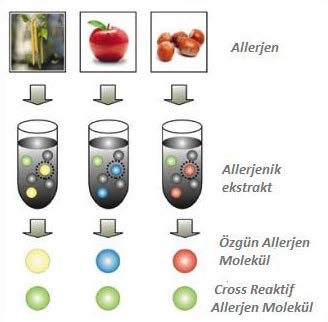
Figure 1. Specific allergen components from a basic allergen source.

What can allergen components tell us?

Allergen components are proteins that have structural similarity and are grouped under different protein families. The reason for the sensitivity developed against these components, which are found in different amounts in the sources and have different stability is due to their common group characteristics. Depending on the properties of these proteins, the sensitization developed by the patient produces different results. Some allergen components are specific and some are cross-reactive.

What Other Contribution Does Molecular Allergology Provide?

With Molecular Allergology, specific allergen components can be produced from a basic allergen source for advanced diagnosis. Sensitization to these components is measured individually in a separate test and at a precise molecular level it is determined which component the patient is sensitive to. This information provides the basis for the highly sensitive diagnosis of allergy. In Molecular Allergology, extract-based tests are used together with component-specific analyses.

While the extract gives the answer to which allergen source the patient is sensitizing to, the allergen components provide vital information about risk, specificity and cross-reaction.

1) Determines the risk of clinical reaction

Molecular Allergology draws conclusions on the risk associated with sensitization. Sensitization to stable allergen components can elicit local as well as systemic reactions, while sensitization to unstable components is mainly associated with local reactions.ISAC: Moleküler Alerji Paneli - 14.05.2018 - BİLİMSEL BÜLTENLER - Biruni Laboratuvarı - 0850 241 77 88

Şekil 2. Risk levels relevant with common allergen proteins

Specific components – provide unique clues to reveal sources of allergies.

Each allergen source typically contains both specific and cross-reactive allergen components. Specific allergen components are almost uniquely related to their source and are only present in small amounts in a limited number of closely related species. Each allergen source may contain one or more specific allergen components. Sensitization to any of these indicates a true sensitization in the person; this means that the relevant allergen source is the primary cause of clinical symptoms.

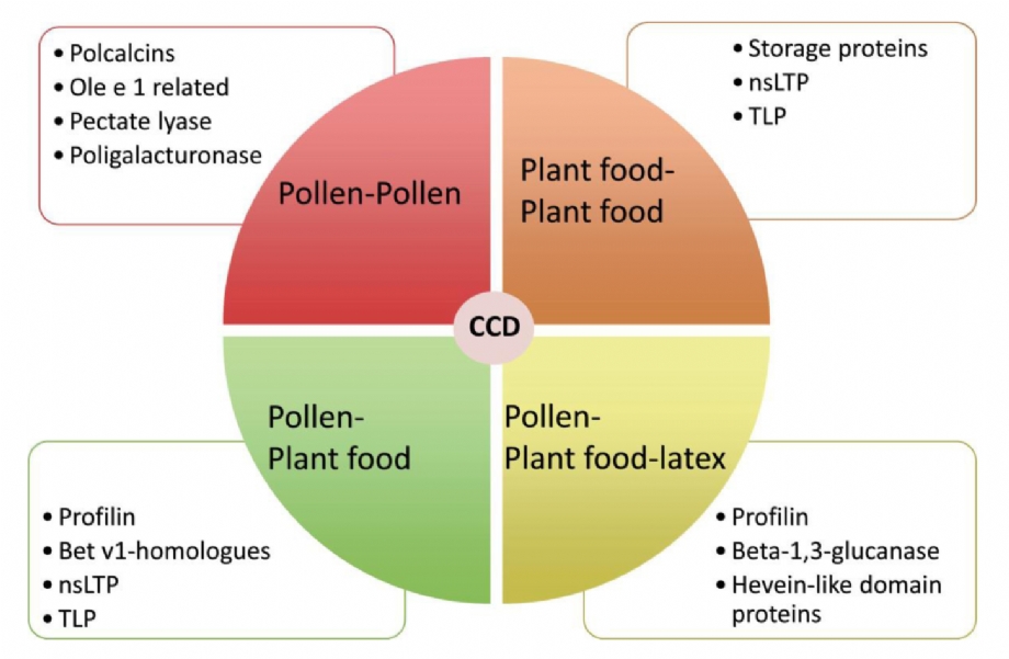
2) Explains symptoms due to cross-reaction

Symptoms elicited by cross-reacting antibodies can be distinguished from those caused by true sensitization, which is important for patient management and appropriate avoidance advice. Where only cross-reaction sensitization is detected, it is necessary to find the primary sensitizer.

Identification of cross-reactive components

It is important to identify the exact trigger of the allergy before starting treatment. However, patients; Cross-reactivity of allergens is common in skin tests and clinical tests such as extract-based antibody tests. Analysis of allergen sources and components of each of the most important pan-allergens allows rapid and accurate identification of the exact triggers of allergy symptoms.

The cross-reaction may be exemplified by birch pollen-related food allergy, a syndrome that affects many birch pollen allergy sufferers. The molecular reason underlying the cross-reaction is that patients with birch pollen allergy have specific IgE antibodies specific to the Bet v 1 component. Bet v 1 has structural similarity to related proteins in many foods, such as soy and peanuts. In this way, the patient’s IgE antibodies to Bet v 1 birch cross-react with these related proteins in soy or peanut.

ISAC: Moleküler Alerji Paneli - 14.05.2018 - BİLİMSEL BÜLTENLER - Biruni Laboratuvarı - 0850 241 77 88

Figure 3: PR-10, Protein- is sensitive to heat and digestion. Cooked foods are often tolerated. It is mostly associated with local symptoms such as OAS. It is related with allergic reactions to pollen, fruits and vegetables.

3) Selection of Appropriate Therapy – Contributes to identifying the right patients for Specific Immunotherapy (SIT).

Determination of sensitization to specific allergen components is necessary for successful Specific Immunotherapy. In the light of these findings, the outcome of the treatment to be applied in patients who develop a true sensitization against the component extracted from the relevant allergen source will be positive. SITs are most likely to be successful when the patient is primarily sensitized to the main components of the allergen extracts. Only molecular allergy diagnoses can offer such in-depth information. The most appropriate treatment can then be selected and patients are relieved of the stress of avoiding unnecessary allergens or ineffective SITs.

ISAC: Moleküler Alerji Paneli - 14.05.2018 - BİLİMSEL BÜLTENLER - Biruni Laboratuvarı - 0850 241 77 88

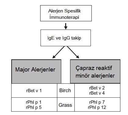
Figure 4 : Recombinant allergen-based diagnostic tests and specific immunotherapy specific to birch and meadow grass allergen

Protein Stability and Amount

Food allergen components show different stability to heating and digestion, and their content in the allergen source may vary. Both stability and quantity are reflected by the protein family to which the component belongs. Therefore, it is possible to assess the risk associated with sensitizations by knowing the patient’s sensitization profile and to which family the identified components belong.

ISAC: Moleküler Alerji Paneli - 14.05.2018 - BİLİMSEL BÜLTENLER - Biruni Laboratuvarı - 0850 241 77 88

Figure 5 : Immunoglobulin E (IgE) levels of allergen molecules based on structural similarity in an allergen family.

a. 2S albumıns (fındık, bakliyat ve tohumlarda stabil depolama proteinleri) arasında değişken, sınırlı çapraz reaktivite.

b. Bet v 1-PR-10 variable cross-reactivity among homologous food allergens.

c. High cross-reactivity (in pollen, latex and foods) due to the strongly preserved and similar structure of the Profilins

For example, in egg allergy, Gal d1 (ovomucoid) is the main allergen and is used as an indicator of allergic reaction severity. Sensitizations to the heat-sensitive components Gal d2 (ovalbumin), Gal d3 (conalbumin), and Gal d4 (lysozyme) are associated with symptoms that occur only with consumption of raw or lightly cooked eggs. Ovalbumin is used in vaccines and lysozyme is used as a preservative, so patients sensitive to these ingredients may have a reaction to drugs or food products containing the relevant ingredient. Similarly, the reaction to Bos d8 (casein) indicates a strong allergy to milk and dairy products. Casein is often used as an additive, so casein sensitivities can cause sensitivities to many different foods, such as chocolate or potato chips. Bos d (lactoferrin), Bosd4, Bos d5 and Bos d6 components are heat sensitive and sensitivities to these components are mainly associated with reactions to fresh milk. Antibodies to Bos d6 (bovine serum albumin) can also cause a reaction to beef.

ISAC: Moleküler Alerji Paneli - 14.05.2018 - BİLİMSEL BÜLTENLER - Biruni Laboratuvarı - 0850 241 77 88

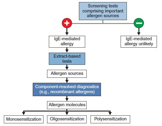
Figure 6 : Serological tests for the diagnosis of allergies

ImmunoCAP ISAC (Immuno Solid-phaseAllergenChip)

ISAC: Moleküler Alerji Paneli - 14.05.2018 - BİLİMSEL BÜLTENLER - Biruni Laboratuvarı - 0850 241 77 88

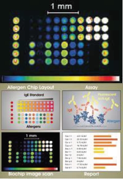
Detection of IgE antibodies in serum plays an important role in the diagnosis of type I allergies. The currently used extract-based method of specific IgE antibodies, ImmunoCAP-FEIA, is the gold standard for allergy diagnosis. However, while this test only allows the simultaneous determination of a limited number of allergens, Immuno CAP ISAC enables the simultaneous detection of 112 allergen components from 48 different main allergen sources. This system is array-based, biochip technology and is the most advanced in vitro diagnostic test for the measurement of specific IgE antibodies for allergen components. The ISAC test facilitates the identification of a comprehensive and specific IgE antibody profile.

ImmunoCAP ISAC is a semi-quantitative test and results report specific IgE antibody levels in ISAC Standard Units (ISU) within the 0.3 – 100 ISU-E measuring range.

ISAC Advantages:

Simultaneous analysis of 112 allergenic proteins allows us to fully and quickly identify the allergens causing the patient’s clinical condition from a single blood sample. The very wide range of allergenic protein studies makes it possible to highlight unexpected sensitizations and/or rule out others. As a result, the analysis allows to obtain an individual sensitization profile with improvement in diagnosis, leading to an individualized treatment. While all this helps improve the patient’s quality of life, the economic cost of analysis is much less than analyzing individual specific IgEs. Most allergic patients test positive to numerous allergens, and the true cause of symptoms may not be determined due to an uncertain medical history regarding the role of different allergens and reactions.

ImmunoCAP ISAC in the diagnosis and treatment of these patients;

  • It sheds light on the true sensitization profile of susceptible patients.
  • It presents the potential risk for severe food-related reactions.
  • It determines the IgE antibody profile in patients with inadequate response to treatment.

Under favour of cross-reacting proteins, ImmunoCAP ISAC can provide information on hundreds of allergen components in addition to the 48 main allergen sources from which the proteins are derived. ImmunoCAP ISAC can reveal unexpected sensitivities or help rule out allergy by providing IgE results for a wide array of allergens. As a result, effective and optimized treatment principles for patients can be initiated earlier and the quality of life is increased while ensuring patient health.

Cross-reacting allergen components are more widely distributed and can be shared among a wide range of allergen sources. Due to their high structural similarity, they can cause cross-reactivity of the IgE antibody.

Components from different protein families produce symptoms of varying severity. Thus, molecular profiling can determine whether a patient has a low or high risk of serious systemic reactions such as anaphylactic shock. Patients at risk of life-threatening reactions can then be advised on allergen avoidance and appropriate measures to be taken in an emergency. For example, if a patient is sensitive to allergens in the profilins family, milder symptoms can be expected overall. Patients who are sensitive to allergens belonging to the family of storage proteins have a high risk of life-threatening systemic reactions. In addition, differences in the heat resistance of protein families play an important role in food allergies.

The prevalence of oral allergy syndrome (OAS) is increasing. It is currently reported to affect approximately 2% of the population in primary care practices in the UK. Although it can often be diagnosed by clinical history, the need for confirmatory testing is increasing for patients with typical oral symptoms after eating raw fruits or vegetables, or sometimes raw nuts. The overlap of symptoms between patients with oral allergy syndrome and patients with nut allergy is an area that often requires more precise definition using available tests to inform patient management and stratify risk. Testing multiple recombinant proteins with ISAC is particularly useful in elucidating patients’ reaction to PR-10 and/or profilin proteins (combined low risk of clinical reaction only) or lipid transfer protein or peanut storage proteins (higher risk of severe reaction).

ISAC Indications:

  • Improving diagnosis in polysensitized patients.
  • To prevent diagnostic errors, especially in patients who do not have a clear correlation between the positive results of conventional allergy tests and symptoms.
  • Preventing therapeutic errors that may occur in terms of Allergen Specific Immunotherapy.
  • To evaluate cases with an inconsistent clinical history of allergy and/or inadequate response to treatment.
  • Evaluation of patients with a history of idiopathic anaphylaxis detecting unexpected sensitivities. ISAC ALLERGEN PROTEIN FAMILIES

ImmunoCAP ISAC provides a large amount of allergen-specific IgE antibody information in a single step. Thanks to cross-reacting proteins, ImmunoCAP ISAC can provide information on hundreds of allergen sources in addition to the 48 sources from which the proteins are derived.

Please Click to overview the ISAC Immunocap Cross-Reaction Map

Storage proteins

  • They are heat and digestion resistant proteins, so they also react with the cooking of foods.
  • They cause more severe systemic reactions in addition to OAS.
  • Proteins found in nuts and seeds as source material for the development of new plants.


Profilin (1)

  • These proteins are sensitive to heat and digestion. Cooked foods are often tolerated.
  • Although rarely associated with clinical symptoms, it may cause local or even severe reactions in some patients.
  • Profilins are present in all pollen and food plants.


PR-10 protein, Bet v 1 homologue(1)

  • Many of the PR-10 proteins are sensitive to heat and digestion. Cooked foods are often tolerated.
  • It is mostly associated with local symptoms such as OAS.
  • It is associated with allergic reactions to pollen, fruits and vegetables.


Polcalcin (Calcium-binding proteins) (2)

  • It is a marker that does not take place in plants used as food, that determines cross-reactivity to pollen.


LTP (non-specific Lipid Transfer Proteins, nsLTP)(1)

  • They are heat and digestion resistant proteins, so they also react with the cooking of foods.
  • They are usually associated with systemic reactions in addition to OAS (Oral Allergy Syndrome).
  • It is associated with allergic reactions to fruits and vegetables in the regions where peach and its derivatives are grown.


CCD (2)

  • It is a marker that determines the sensitivities developed against cross-reactive carbohydrates.
  • Although it is very rare to cause allergic reactions, pollen containing CCD, plants used as food, pests and poisons can cause positive IVD results.


Lipocalin (3)

  • They are important allergens that are constantly found in animals.
  • It is a component that shows limited cross-reactivity between animal species.


Parvalbumin (3)

  • These proteins are resistant to heat and digestion. They also cause a reaction in cooked foods.
  • They cause more severe systemic reactions in addition to OAS.
  • The main allergen is found in fish and is a marker that determines cross-reactivity between different fish species and amphibians.


Serum albumin (3,4)

  • These proteins are very sensitive to heat and digestion.
  • Animals inhabit different biological fluids and regions. eg. cow’s milk, blood, meat and epithelium.
  • They cause cross-reactions between different common mammalian species. For example: Cat-dog and pig

ISAC IMPORTANT ALLERGEN COMPONENTS

Gal d 1, Ovomucoid (egg white / glair) (3)

  • Ovomucoid IgE antibodies are associated with persistent egg allergy and are generally intolerable both raw and cooked.


Tropomyosin (3)

  • These proteins are resistant to heat and digestion. They also cause a reaction in cooked foods.
  • They cause more severe systemic reactions in addition to OAS.
  • It is actin-binding proteins in muscle tissues and is a marker that determines cross-reactivity between shellfish, mites and cockroaches.


Ara h 1, 2, 3, 6, 8 and 9 (peanut) (3)

  • Intermediate h 1, 2, 3, 6 and 9 (LTP) IgE antibodies cause peanut-induced systemic reactions in addition to OAS.
  • Intermediate h 8 (PR-10) IgE antibodies generally cause milder local symptoms such as OAS. It is often associated with birch-related sensitivities.


Gly m 4, 5 and 6 (soy) (3)

  • Patients allergic to soybean often have Gly m 5 and Gly m 6. Gly m 5 & Ara h 1 and Gly m 6 & Ara h 3 IgE antibodies. Cross-reactivity and clinical reactivity may be seen due to IgE of the storage proteins found in these legumes.
  • Gly m 4 (PR-10) IgE antibodies are usually associated with local symptoms such as OAS and arise from birch sensitivity. However, some cases of severe reactions to Gly m 4 have also been reported. eg. during birch pollen season, often combined with exercise and intake of less processed soy beverages.


Alt a 1 (Alternaria) (3)

  • Alt a 1 is the main allergen of Alternaria, which is associated with the development of asthma.


Tri a 19, Omega-5 gliadin (wheat) (5,6,7)

  • Omega-5 gliadin (Tri a 19) IgE antibodies in adults are associated with reactions triggered by exercise or the use of NSAIDs due to wheat ingestion.
  • Omega-5 gliadin (Tri a 19) IgE antibodies are associated with an increased risk of immediate reactions to wheat in children.


ISAC: Moleküler Alerji Paneli - 14.05.2018 - BİLİMSEL BÜLTENLER - Biruni Laboratuvarı - 0850 241 77 88


A New Metabolic Organ: “Intestinal Microbiota”

Since the moment which we have born there are many microorganisms accompanying us in our body. The human body contains a microbial population, mostly bacteria, including fungi, viruses and protozoa. Bacteria are generally known as disease-causing pathogens. However, humans live in a symbiotic balance with bacteria. It should not be forgotten that we need bacteria and their beneficial effects to stay healthy.


Many different microbial populations reside in the human body, mostly bacteria, including fungi, viruses, and protozoa. This population contains 10 times more microbial cells than human cells and 150 times more genes than the human genome. This ecological community formed by commensal, symbiotic and pathogenic microorganisms that share our body is called “microbiota”. “Microbiome” is defined as the total genome of microorganisms living in this environment. (1)

The human microbiota has been colonized in the skin, genitourinary system, respiratory system, and most often in the gastrointestinal tract. The gastrointestinal tract contains the richest microorganism community in our body, due to its surface area of approximately 200m² and rich nutrients for microorganisms. In healthy individuals, the microbiota includes a large number and variety of microorganisms. It begins to form immediately after birth. It varies according to nutrition, genetics, age and geographic region. Bacteria count is 10⁹ ml/saliva in the mouth, 102-4 g/material in the stomach, 10¹¹ g/material in the colon and 10¹² g/material in the stool. The mode of birth, diet, and genetic factors affect the microbiota in infants. Intestinal microbiota may change after treatment applications such as infections and antibiotic use (2)

Dietary content plays an important role in the change of intestinal flora. A diet rich in fiber foods facilitates the proliferation of Firmicutes bacteria such as Eubacterium rectale, Eubacterium halli, Rumicoccus bromii (Picture 1).


Picture 1: Effect of nutrition on intestinal microbiota (Flint et al.) WK: Wheat-containing food BS: Fiber-containing food EW: Protein-containing food

Intestinal microbiota; It plays a very complex and active role on the physiological, metabolic and immune system in our body. Intestinal bacteria control the necessary metabolic processes by acting as energy carriers or releasing immune-modulating substances. For this reason, the intestinal microbiota is now defined as a new “metabolic organ” (3).

Commensal intestinal bacteria;

  • It breaks down indigestible foods and makes them useful for the body.
  • Supports the digestion of complex carbohydrates and fibers.
  • It prevents the reproduction of pathogenic bacteria.
  • Contributes to the production of vitamins B1, B2, B6, B12 and K.
  • Contributes to the detoxification of toxins and wastes.
  • They ferment carbohydrates and proteins taken with food and turn them into short-chain fatty acids such as lactic acid, butyric acid, acetic acid, and gases such as hydrogen and carbon dioxide.
  • Short-chain fatty acids are an energy source for intestinal mucosal cells.
  • Short-chain fatty acids contribute to proper intestinal peristalsis.
  • Butyrate provides a strong anti-inflammatory activity by inactivating Nuclear Factor Kappa (Factor NF-kB) transcription and IL-8 production. It is accepted that Firmicutes bacteria, especially Faecalibacterium prausnitzii, play the most active role in butyrate production. These bacteria make up 5-15% of the intestinal flora. Acute phase reactant proteins such as alpha 1 antitrypsin and calprotectin are responsible for inflammatory irritation in the intestinal mucosa.

A decrease in F. prausnitzii correlates with an increase in the degree of inflammation.

  • Colon epithelial cells in healthy individuals are covered with a protective mucous layer. Bacteria of the genus Verrucomicrobia contribute to immune modulation by promoting mucosal production, especially from Akkermansia muciniphila goblet cells. When the mucosal layer is damaged or mucin production is insufficient, pathogens, pollutants and allergens come into direct contact with the mucosal cells and cause inflammation. (4,5)

DYSBIOSIS

Intestinal microbiota may change due to chronic gastrointestinal diseases as antibiotic use. Disturbance in the intestinal microbiota balance is defined as “dysbiosis”. It has been shown that there is an increase in intestinal permeability, a change in the production of short-chain fatty acids, and a decrease in colonic resistance when the microbiota balance is disturbed. A decrease in Firmicutes strains and an increase in Proteobacteria species such as Salmonella, Shigella, Klebsiella, Proteus, Escherichia coli are associated with various diseases.

Sulfate-consuming bacteria lead to the production of hydrogen sulfide (H2S), paving the way for the development of intestinal diseases. H2S is a toxic metabolic product that damages the intestinal epithelium, resulting in cellular atypia. Bilophilia wadworthii, Desulfomonas pigra and Desulfovibrio piger species are bacteria that play an important role in H2S production. Clostridium species, which are obligate anaerobes, are pathogenic bacteria due to their immune modulatory effects and increasing IL-10 production. Toxin-producing origins of bacteria of the genus Clostridium, in particular, are detected in patients with autistic spectrum, often causing intestinal and extra-intestinal autistic complaints.

Haemophilus and Fusobacteria species, which are known as vepathogens in the respiratory tract mucosa, can also be detected in the intestines. Studies show that these pathogenic strains are associated with chronic inflammatory bowel diseases, colorectal carcinomas and appendicitis. As molecular-genetic studies in stool increase, this relationship will be better defined (6,7).

RELATIONSHIP OF DYSBIOSIS AND CLINICAL SYMPTOMS

Pretty valuable results have been obtained with detailed analyzes of the intestinal microbiota. The ideal organization of the intestinal flora population is one of the main elements of a healthy physiological life. When the intestinal microbiota is defined personally, solutions such as diet regulation according to the presence of increased or decreased origins and the use of appropriate prebiotic probiotics can be applied.


1. Obesity and Metabolic Syndrome

In healthy individuals, the Firmicutes/Bacteroidetes ratio usually ranges from 1:1 to 1:3. In overweight people, this ratio varies from 3:1 to 25:1. It has been shown to reach a ratio of 200:1 in some overweight individuals (8 ,9).

Another consequence of obesity is a significant decrease in the amount of Faecalibacterium prausnitzii belonging to the firmicute genus. F.prausnitzii is one of the 3 most common bacteria in the intestinal flora. It produces butyrate. Butyrate supports the development of the intestinal mucosa. Butyric acid salts inhibit the transcription of Factor NF-kB and inhibit the release of additional chemokines and interleukin 8. In obese, hsCRP and interleukin 6 levels are significantly increased, at the same time the amount of F.prausnitzii is decreased. In these patients, when the amount of F.prausnitzii is increased, the mucosa can be protected and inflammatory reactions can be reduced (4).

A. muciniphila species that contribute to the production of mucus produced from goblet cells are also frequently reduced in overweight individuals. Mucus covers the intestinal epithelial cells and forms a barrier that protects them from chemical and mechanical effects. It has been shown that the amount of Akkermansia muciniphila is significantly reduced in people who are fed a high-fat diet. The number of bacteria can be partially increased by adding prebiotics containing oligosaccharides to the diet of these people. In animal experiments, the positive effects of A. muchiniphila supplementation on weight loss, development of mucosal layer and reduction of fasting blood glucose and insulin resistance have been shown. It has been reported that similar results have been obtained in humans (10).

2. Intestinal Inflammation

Irritable bowel syndrome is a common, long-lasting clinical picture that is seen in many people and manifests itself with attacks. Recent studies have shown that the amount of F. prausnitzii is reduced by approximately 30% in people with irritable colon complaints and Crohn’s disease. Considering that F. prausnitzii species play the most important role in the production of butyrate, which has an anti-inflammatory effect, and the inhibitory effect of butyrate on Factor NF-kB and IL-8, this decrease negatively affects the anti-inflammatory effect on the mucosa (4,6)

Campylobacter species are isolated in approximately 70% of children newly diagnosed with Crohn’s disease. For this reason, when Campylobacter species are isolated in the stool, there is a growing debate that administration of probiotics will reduce pathogenic bacteria (11).

Leaky gut syndrome is a clinical pattern suggested to be closely related to the intestinal microbiota. The amounts of A. muchinophilia and F. prausnitzii were decreased in these individuals. Intestinal cells are like bricks lined up next to each other. There are “tight connections” between them, which we can call cement between bricks. Thus, undesirable substances cannot pass out of the intestine (ie inside the body) from here, they remain in the intestine and are excreted from the large intestine. In order for the shape of the intestinal cells and the connection between the cells to be healthy, the cells must be taut, which is when the intestines have enough energy to stay taut. F. prausnitzii species use dietary polysaccharides to form short-chain fatty acids. Energy is also produced from short-chain fatty acids. Just on the outer surface of the intestine, there are immune system cells that examine the substances passing through the intestine. When there is an excessive passage through the gut, these immune system cells become active and initiate a reaction, but this reaction is too small to cause disease.

This is called low-level inflammation. Low-level inflammation does not end as long as intestinal permeability continues, which causes all the body’s energy to be used by immune system cells in a long time. Therefore, sufficient energy cannot go to other organs in need, and some problems begin to occur in these organs. Another bad side of hyperpermeable bowel syndrome is that unwanted substances that enter the body go to the weak tissues of the body and accumulate there, and in the long term, the immune system attacks these tissues, causing autoimmune diseases (12,13).

3. Intestinal Tumors and Intestinal Cancers

In addition to other known carcinogenic effects that acids, especially hydrogen sulphate, increase atypical cell growth, cause mucosal irritation and predispose to colorectal cancer. Sulfate producing bacteria are Desulfomonas piger, Desulfovibrio piger and H2S producing Clostridium species. Pro-prebiotic therapies can be applied when an increase in the number of sulfate-producing bacteria is seen in the stool microbiome analysis.

It has also been shown that intestinal microbiota changes in intestinal tumors. In these individuals, the amount of F. prausnitzii is frequently reduced to an undetectable (13).

4. Arthritis

Bacterial imbalances are detected in the intestinal microbiome of patients with rheumatoid arthritis, paralleling the development and progression of the disease. For example, Prevotella copri is beneficial for the immune and digestive system when it is within physiological limits in the intestinal microflora. However, it has been reported that the amount of Prevotella copri and other prevotella species is significantly increased in patients with rheumatoid arthritis. It is suggested that this situation prevents other beneficial bacteria from reproducing and performing their functions (14).

5. Autism

Genetic factors play a big role in autism. However, many other factors can also cause the development of the disease. Many intestinal diseases accompany clinical complaints including the autistic spectrum. Studies show that the use of antibiotics not only eases intestinal complaints, but also increases other symptoms of autism. It is suggested that intestinal microflora also contributes to brain development through the brain-gut axis. It has been reported that the deterioration of gut biodiversity not only leads to the development of autism, but also increases the severity of symptoms. Toxin-producing Clostridium species are found to be increased in children with autism. More Clostridium species are isolated in children with autism than in the control group with normal neurological development. However, it is not yet fully understood how the excess of Clostridium species plays a role in the onset and development of autism. When toxin-producing strains of Clostridium species are detected in the stools of children with autism, appropriate probiotic use is recommended (7,15).

6. Alzheimer Disease

In a recent study, it was shown that the amount of F. prausnitzii decreased by 100% in the intestinal microflora of Alzheimer’s patients (n=52). In addition, inflammation indicators such as calprotectin and antitrypsin were found to be increased in 87.5% of the stools of these patients. hsCRP values are high in 91% of these patients. These data indicate the presence of a systemic inflammation in the body, and it is suggested that a decrease in the amount of F. prausnitzii may be the cause of this inflammation (16). As a result, with personalized advice and personalized treatment approaches are possible by determining the personal gut microbiota.

References & Sources

  1. Turnbaugh, P.J.; Ley, R.E.; Hamady, M.;Fraser-Liggett,C.M.; Knight, R.; Gordon,J.I. (2007). “The Human Microbiome Project”. Nature 449: 804-81O.
  2. Bull M.J., Plummer N.T.. Part 1: The Human Gut Microbiome in Health and Disease. in: lntegrative Medicine: A Clinician’s Journal 13(6), S. 17-22, 2014
  3. Jandhyala, S. M. et al: Role of the normal gut microbiota. in: World J Gastroenterol 21(29), S.8787-8803,2015
  4. Miquel, S. et al.: Faecalibacterium prausnitzii and human intestinal health. in: Curr Opin Microbiol. 16(3), S. 255-261, 2013
  5. Everard A., et al.: Cross-talk between Akkermansia muciniphila and intestinal epithelium controls diet-induced obesity. in: PNAS 110(22), S. 9066-9071, 2013
  6. Ramezani, A. et al.: The Gut Microbiome, Kidney Disease, and Targeted lnterventions. in: JASN 25(4), S. 657-670, 2014
  7. Song, Y. et al.: Real-Time PCR Quantitation of Clostridia in Feces of Autistic Children . in: AEM 70, S.6459-6465,2004
  8. The NIH HMP Working Group et al.: The NIH Human Microbiome Project. in: Genome Res. 19, S. 2317-2323, 2009.
  9. Kasai et al. Comparison ofthe gut microbiota composition between obese and non-obese individuals in a Japanese population, as analyzed by terminal restriction fragment length polymorphism and next-generation sequencing BMC Gastroenterology (2015) 15:100 DOI10.1186/sl 2876-015-0330-2
  10. Everard, A. et al.: Cross-Talk between Akkermansia muciniphila and lntestinal Epithelium Controls Diet-lnduced Obesity. in: PNAS 110(22), S. 9066-9071, 2013
  11. Deshpande, N. P. et al.: Comparative genomics of Campylobacter concisus isolates reveals genetic diversity and provides insights into disease association. in: BMC Genomics 14, 585, 2013
  12. Michielan, A. et al.: lntestinal Permeability in lnflammatory Bowel Disease: Pathogenesis, Clinical Evaluation, and Therapy of Leaky Gut. in: Mediators of lnflammation, 2015, 628157
  13. Nava G.M. et al.: Abundance and diversity of mucosa-associated hydrogenotrophic microbes in the healthy human colon. in: The iSME Journal 6(1), S. 57-70, 2012
  14. Seher, J. U. et al.: Expansion of intestinal Prevotella copri correlates with enhanced susceptibility to arthritis. in: elife, 2, e0l 202, 2013
  15. Smith, P.A.: Brain, meet gut. in: Nature 526, S. 312-314, 2015
  16. Leblhuber, F. et al.: Elevated fecal calprotectin in patients with Alzheimer’s dementia indicates leaky gut. J Neural Transm (Vienna) 122(9) S. 1319-1322, 2015
  17. Mandal, S. et al.: Analysis of composition of microbiomes: a novel method for studying microbial composition. in: MEHD 26, S. 27663-27670, 2015